incisive canal radiograph
Contribs created this work entirely by myself color006400Contribs. This canal may also be referred to as the incisive canal.
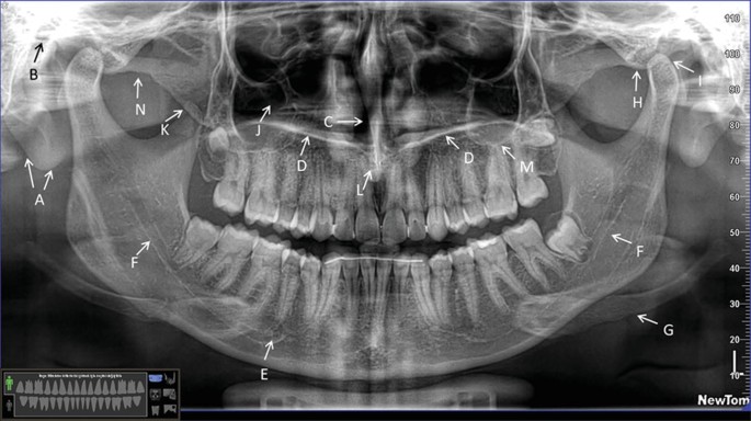
Panoramic Radiographic Anatomy Springerlink
Original uploader was DRosenbach at enwikipedia.

. It connects the inferior nasal cavity with the superior oral cavity opening at the incisive foramen posterior to the central maxillary incisor teeth. Le Courrier de Bellechasse 1937-1941 1957-1960 1962-1963. Symmetry When evaluating radiographs first consider symmetry.
National Center for Biotechnology Information. Periapical radiograph panoramic and CBCT are needed to assess the lesion with better precision and limits radiation. 30 November 2010 1346 UTC Source Original text.
Die Bewertungen umfassten 1 den mesiodistalen Durchmesser 2 den labiopalatalen Durchmesser 3 die Länge des Schneidekanals 4 die Form des Schneidekanals und 5 die Breite des Knochens vor dem Foramen incisive. Panoramic radiographs can be used for visualization of the mental foramen and a potential anterior looping but not for locating the mandibular incisive canal. On periapical x-ray images the incisive foramen is located in the midline between the roots of the central incisors.
Its appearance is quite variable due to normal anatomic variation and due to the operators angulation of the x-ray beam. 323 098 mm. The aim of this study was to investigate the presence of a MIC in panoramic radiographs OPGs.
Popularly known as nasopalatine canal is a radiolucent tube shaped area located in between the maxillary central incisors. 64 shows apical root lateral displacement secondary to the cystic lesion. Le Courrier de Papineau 1961 1965-1966.
Panoramic radiographs can be used for visualization of the mental foramen and a potential anterior looping but not for locating the mandibular incisive canal. One thousand forty-five OPGs were randomly chosen from patient population. It represents the anterior continuation of the mandibular canal.
An anatomical variation to be considered is the anterior looping of the mental nerve in 11 of images. The differential diagnosis for incisive canal cyst includes medial enlarged nasopalatine duct central giant cell granuloma central incisor root cyst. Usually only the inferior border of the orbit is visible over the panoramic radiograph Incisive canal.
Incisive Fossa Radiograph - ultimate radiology incisive canal cyst nasopalatine duct cyst canine radiographs ppt figure powerpoint presentation free download id 2282480 maxilla bone anatomy features of an ideal panoramic radiograph practical panoramic imaging Home Incisive Fossa Radiograph Incisive Fossa Radiograph Lisa. An incisive canal was identified in 93 of the cases with good visibility in 22 of the cases. The incisive foramen also known as nasopalatine foramen or anterior palatine foramen is the oral opening of the nasopalatine canal.
It contains the descending palatine artery and the nasopalatine nerve. It is seen on both intraoral radiographs and extraoral radiographs. It can be single or multiple.
Le Courrier 1949 1951. Die mittlere Breite des Foramen labiopalatal und mesiodistal betrug 312 094 mm bzw. Panoramic radiographs can be used for visualization of the mental foramen and a potential anterior looping but not for locating the mandibular incisive canal.
Citation DOI article data. The mandibular incisive canal MIC is a small bony channel located in the interforaminal region. Mean sd vertical diameter buccolingual diameter and inner diameter of the incisive canal were 47 11 37 07 and 11 03 mm respectively.
This root displacement is absent in a normal incisive canal. The nasopalatine canal presents as a vertical radiolucent band between the roots of the maxillary central incisors superiorly to the Post topics. The maxillary incisive canal runs through the maxilla in the midline.
64 Plain film radiograph demonstrating apical root lateral displacement secondary to an incisive canal cyst. An incisive canal was identified in 15 of the images with good visibility in only 1. Incisive canal cysts are treated with complete surgical removal by a palatal approach with the palatal flap.
Periapical radiograph depicting the junction of the mandibular canal and the mandibular incisive canal near the mental foramen. This file includes issues of the following newspapers. Panoramic radiograph PAN is an extraoral radiographic technique widely used by many implantodontists and oral and maxillofacial surgeons.
To verify its existence for preoperative planning purposes cross-sectional imaging modalities HR-CT or spiral tomography should be preferred. Incisive foramen is the opening of the incisive canal located immediately behind the maxillary central incisors. However the reliability of measurements obtained by this method is low due to distortion and.
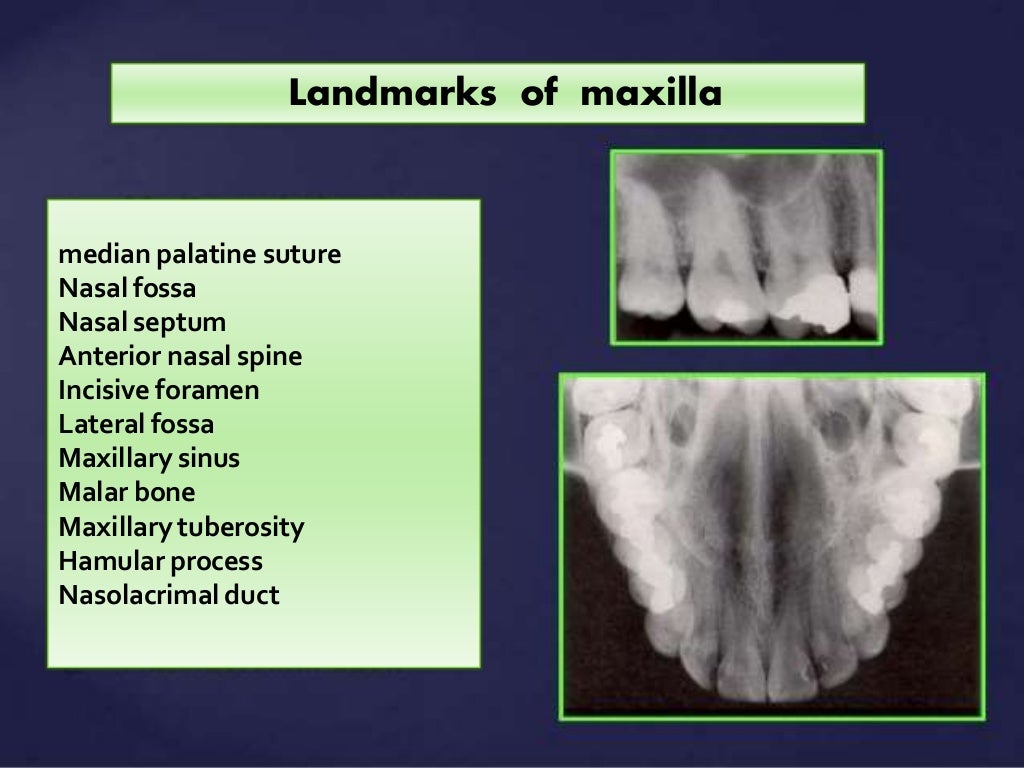
It is located in the maxilla in the incisive fossa midline in the palate posterior to the central incisors at the junction of the medial palatine and incisive sutures. The mandibular incisive canal MIC is the anterior extension of the mandibular canal and its presence is of interest in surgical procedures in the chin region.
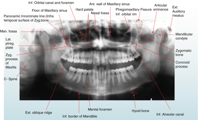
Panoramic Radiographic Anatomy Springerlink

Intraoral Radiographs Identifying Normal Anatomy Today S Veterinary Practice

Mouth Incisive Canal Cyst Professional Radiology Outcomes

Anatomical Landmarks Of Panoramic Radiographs With Ppt Lecture Note For Download Lecture Notes In Dental Assistant Study Dental Hygiene School Dentistry

Automatic Visualization Of The Mandibular Canal In Relation To An Impacted Mandibular Third Molar On Panoramic Radiographs Using Deep Learning Segmentation And Transfer Learning Techniques Oral Surgery Oral Medicine Oral Pathology

Fundamentals Of Radiographic Interpretation For The Dentist Pocket Dentistry

Panoramic Radiograph Image A Axial B And Oblique Sagittal C Ct Download Scientific Diagram

Dentaltown Where The Dental Community Lives Denti Dentista Odontoiatria

Pdf The Evaluation Of Visibility Of Mandibular Anatomic Landmarks Using Panoramic Radiography Semantic Scholar

Analysis Of Dental Radiographs And Cbct Studies Springerlink

Measurement Of Incisive Foramen Blue Line Nasal Foramen Red Line Download Scientific Diagram
Normal Radiographic Anatomical Landmarks

Opg Showing Incisive Foramen And Mental Foramen Download Scientific Diagram

6 Essentials Of Dental Radiographic Analysis And Interpretation Pocket Dentistry

Transient Apical Breakdown

Periapical Radiograph 1 Year After Treatment Bone And Teeth Showing Download Scientific Diagram
Panoramic Radiographic Anatomy Springerlink

Route Of The Incisive Canal Of The Mandible Mic Download Scientific Diagram
![]()
Panoramic Radiograph Showing Mandibular Incisive Canal Arrow Download Scientific Diagram
0 Response to "incisive canal radiograph"
Post a Comment